Close this window to return to the previous window
Close this window to return to the previous window |
|||
|
|||
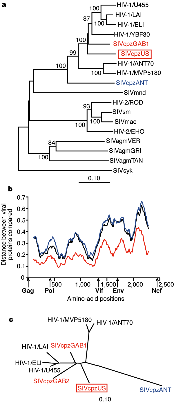
| Figure 1 Phylogenetic analysis of SIVcpzUS. a, Phylogenetic relationship of SIVcpzUS to other primate lentiviruses. The tree was derived by neighbour-joining analysis27 of full-length Pol sequences (trees derived by maximum-likelihood methods28 yielded very similar topologies). Horizontal branch lengths are drawn to scale with the bar indicating 0.1 amino-acid replacements per site. Numbers at each node indicate the percentage of bootstrap samples (out of 1,000) in which the cluster to the right is supported (only values >80% are shown). Other SIVcpz strains closely or more distantly related to SIVcpzUS are shown in red and blue, respectively. b, Diversity plots of concatenated SIVcpz protein sequences depicting the proportion of amino-acid sequence differences between SIVcpzUS and SIVcpzGAB1 (red), SIVcpzUS and SIVcpzANT (blue), and SIVcpzGAB1 and SIVcpzANT (black), calculated for a window of 200 amino acids moved in steps of 10 amino acids along the alignment (available as Supplementary Information). The x-axis shows the amino-acid positions along the alignment. The positions of Gag, Pol, Vif, Env and Nef regions are shown. The y-axis denotes the distance between the viral proteins compared (0.1 = 10% difference). c, Unrooted neighbour-joining tree of partial Pol protein sequences (distances are drawn to scale). | |||
| Note: Figures may be difficult to render in a web browser. In such cases, we recommend downloading the PDF version of this document. | |||